Stan książek
Nasze książki są dokładnie sprawdzone i jasno określamy stan każdej z nich.


Nowa
Książka nowa.
Używany - jak nowa
Niezauważalne lub prawie niezauważalne ślady używania. Książkę ciężko odróżnić od nowej pozycji.
Używany - dobry
Normalne ślady używania wynikające z kartkowania podczas czytania, brak większych uszkodzeń lub zagięć.
Używany - widoczne ślady użytkowania
zagięte rogi, przyniszczona okładka, książka posiada wszystkie strony.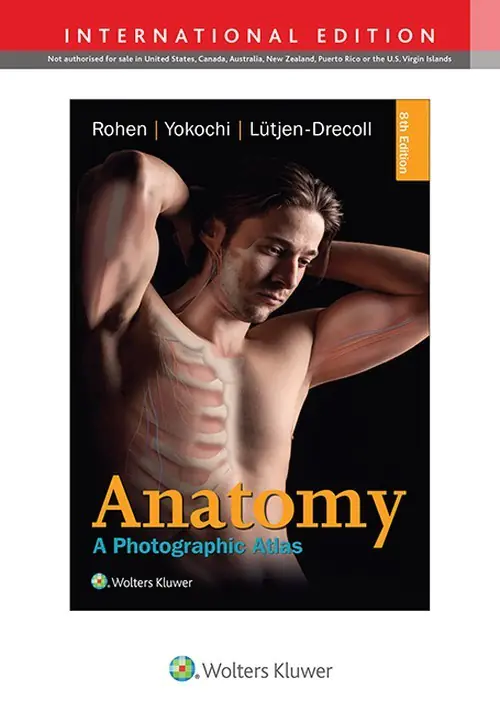
Anatomy: A Photographic Atlas 8e
Masz tę lub inne książki?
Sprzedaj je u nas

This revised eighth edition of "Anatomy: A Photographic Atlas" offers a superb collection of full-color photographs from real cadaver dissections, complemented by schematic drawings and diagnostic images. Unlike traditional atlas illustrations, this reliable resource presents anatomical structures with a greater degree of realism, providing students with a clear depiction of what they will encounter in the dissection lab. The chapters follow the typical order of dissection, organized by region, and systematically introduce topographical anatomical structures in each section. This edition is enhanced with additional clinical imaging, including MRI and CT scans, as well as endoscopic techniques. New graphical elements have been incorporated, highlighting clinically relevant nerve and vessel variations and muscle functions. Many older images have been updated with new high-resolution replacements, and where black-and-white photography was previously used, it has now been substituted with vibrant color imagery.
Wybierz stan zużycia:
WIĘCEJ O SKALI
This revised eighth edition of "Anatomy: A Photographic Atlas" offers a superb collection of full-color photographs from real cadaver dissections, complemented by schematic drawings and diagnostic images. Unlike traditional atlas illustrations, this reliable resource presents anatomical structures with a greater degree of realism, providing students with a clear depiction of what they will encounter in the dissection lab. The chapters follow the typical order of dissection, organized by region, and systematically introduce topographical anatomical structures in each section. This edition is enhanced with additional clinical imaging, including MRI and CT scans, as well as endoscopic techniques. New graphical elements have been incorporated, highlighting clinically relevant nerve and vessel variations and muscle functions. Many older images have been updated with new high-resolution replacements, and where black-and-white photography was previously used, it has now been substituted with vibrant color imagery.
Szczegóły
Dane producenta:


Inne książki tych autorów

Podobne produkty
Może Ci się spodobać
Inne książki z tej samej kategorii

Napisz opinię o książce i wygraj nagrodę!
W każdym miesiącu wybieramy najlepsze opinie i nagradzamy recenzentów.
Dowiedz się więcejWartość nagród w tym miesiącu
880 zł
Dodaj swoją opinię
Zaloguj się na swoje konto, aby mieć możliwość dodawania opinii.
Czy chcesz zostawić tylko ocenę?
Dodanie samej oceny o książce nie jest brane pod uwagę podczas losowania nagród. By mieć szansę na otrzymanie nagrody musisz napisać opinię o książce.
Już oceniłeś/zrecenzowałeś te książkę w przeszłości.
Możliwe jest dodanie tylko jednej recenzji do każdej z książek.
Sposoby dostawy
Odbiór w punkcie (7)
InPost Paczkomat 24/7

14,99 zł
DPD Pickup Automaty i Punkty Odbioru

11,99 zł





Dostawa na adres (7)
GLS U Ciebie - Kurier

14,99 zł
Kurier DPD

14,99 zł





Sposoby płatności
Płatność z góry
Przedpłata

Zwykły przelew
